MORE ABOUT PROTEINS & LAB TECHNIQUE
When you learned how to meaure the concentration of albumin in solutions, and when you studied peroxidase activity, you were learning more than just facts about two particular proteins. Since those two are representative of many others, what you learned about them can be applied to many other proteins. To develop your understanding more fully, and because proteins are so important in so many aspects of biology (medicine, research, biochemistry, industrial applications, etc.), today you'll look more closely at their properties and additional techniques used in studying them. Part of this work involves assays of the peroxidase enzyme. Refer to your previous guidesheet (lab #6) for the details of preparing another enzyme extract and the steps of the basic assay procedure.
Before returning to the peroxidase, though, you'll see some visually dramatic examples of protein behavior in connection with some important lab techniques:
a. effect of pH on proteins in solution
b. effect of an organic solvent on proteins in solution
c. centrifugation as a separation technique
As you see the effects of various manipulations on proteins in tubes, remember that proteins in cells and in biological fluids would be affected similarly. If you could isolate and purify the peroxidase present in your crude enzyme extract, the amount would be too small to see, although the result of this enzyme's activity is readily visible as the color change of solutions. (The beauty of enzymes is that so little accomplishes so much.) Even so, the enzyme molecules in the extract would be affected by these manipulations just as the other proteins are affected.
Wear gloves and splash goggles throughout the work today; handle all solutions carefully, as always. Today be especially careful in handing the 4M HCl (hydrochloric acid, which is in one of the screw cap tubes. If you accidentally spill any of the HCl tell the TA immediately so that he/she can neutralize it. There is a box of NaHCO3 (sodium bicarbonate) for this purpose.
I. Precipitation of milk protein & centrifugation as a separation technique
Milk is a good source of dietary protein, containing about one gram per fluid ounce (30 mL). The major milk protein is called casein. Casein's solubility in water depends on the same aspects of protein structure covered in class and in the text. In order to dissolve in water, protein molecules must be effectively surrounded by "coats of water" (hydration shells). Hydration of the molecules reduces their interaction with each other so that they do not stick together and form precipitates.
A common result of altering the 3-dimensional structure of proteins is precipitation of the casein from the solution. This is what happens when contaminating bacteria in milk produce acid (usually lactic acid) as a byproduct of their metabolism. As the milk's pH decreases, the casein becomes less soluble (as its shape changes), and it precipitates when the casein molecules interact with each other more than with the water molecules. You see this as curdling. In fact, this "spoilage" is used in production of cheeses, yogurt, and other fermented dairy products. The precipitated casein is separated as curds, leaving the fluid portion of the milk as the whey. There are other proteins that remain in the whey; an internet search will provide much interesting information about milk’s composition and the various components found in cheeses, yogurt, etc.
A. Effect of pH. Use a 2 mL pipette and pipette pump to dispense 3 mL of 4 M HCl (hydrochloric acid) into a large plastic test tube (actually, a centrifuge tube). Handle the HCl carefully. Then use the graduated cylinder to add 25 mL of the nonfat milk to the tube. Use the flattened spatula to thoroughly mix the acid and milk. Look carefully to see the curdling (coagulation; formation of precipitate). For the moment set this tube aside in the plastic beaker. Go on to item B, next.
B. Effect of organic solvent. Dispense 9 mL of milk (graduated cylinder) into the other plastic tube. Rinse the cylinder and use it to measure 19 mL of 95% (v/v) ethyl alcohol (FLAMMABLE!). Stir well with the spatula and observe again the formation of a fluffy precipitate.
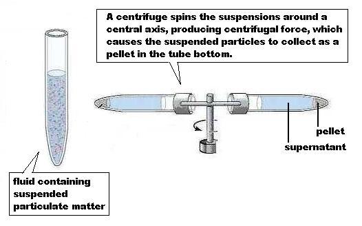
C. Centrifugation. At this point you could recover the precipitated casein in various ways, by simple filtration for example. We'll use centrifugation (see illustration). The principle is simple. If a liquid has particulate material suspended in it, that solid matter can be separated from the liquid by subjecting it to centrifugal force in an instrument called a centrifuge, of which there are many types. The greater the force applied, the smaller the particles that can be forced to collect in the bottom of the centrifuge tube. [Note that material "suspended" in a liquid is not the same as material "dissolved." Suspended material will settle out in time; dissolved material won't.]
Give your two milk samples to the lab instructor to be loaded into the centrifuge. He/she will explain operation of the instrument, balancing of the tubes, and safety. After the short centrifugation period, recover your tubes. You will see the casein "pellet" in the bottom of the tube and the liquid "supernatant" above. Note that the supernatant is transparent; it contains the lactose, minerals, and vitamins of the original milk. Why is one of the two pellets so much larger than the other? (Hint: reread carefully what you did to prepare the 2 tubes.)
Take the acid-precipitated sample to the sink and pour the supernatant into
the sink; the pellet stays in the tube. Put 15 mL of water into the tube
and use the spatula to redissolve the precipitated
protein. By the time all the casein is redissolved,
the new solution contains the protein but very little of the other solutes
originally in the milk; those components were in the supernatant. So, you've
performed a partial purification of the protein. What's more, by redissolving the casein in 15 mL rather than the original
25 mL volume you've also concentrated the protein. Such concentrating of
substances is an important use of centrifugation in addition to separation of
substances.
Although the rotor in this table-top centrifuge spins at about 1000 rpm, there
are centrifuges that will spin much faster and provide much greater centrifugal
force, enough even to separate ribosomes and viruses from biological
fluids.
*Discard the pellets in the trash container and clean both plastic tubes.*
Note that both the acid and alcohol (organic solvent) treatments caused denaturation of the protein. You should be able to explain the molecular details of what happened to the protein molecules in each case.
Bear in mind that what you have seen happen to casein could happen to other proteins as well. Consider also that centrifugation is a very useful technique for separation and concentration of many biologically important substances from complex mixtures: cells, organelles, and molecules.
II. Continuation of study of effects of various factors on enzyme activity - background
Be sure to print a copy of the SETUP TABLE...both part 1 and part 2.
Returning now to peroxidase, in the previous exercise you saw how reaction rate is affected by substrate concentration and by enzyme concentration. Today you will see several other factors that affect reaction rate. Some factors exert their effect by changing the structure of the enzyme molecule.
A. Enzyme extract preparation. Follow the instructions in your previous guidesheet for preparation of an enzyme extract exactly as before. Keep the extract cold in an ice bath as before.
B. The basic assay procedure. The assays are performed as in the previous exercise, except where noted otherwise. The SETUP TABLE shows the volumes of solutions to use today. To perform each assay:
1. Put the 0.2 mL of enzyme extract into one large test tube, and (unless the instructions say otherwise) put the other solutions into a second large test tube. Don't mix up your pipettes; pipeting acid and the enzyme extract with the same pipette would contaminate both solutions and foul the results.
2. Then pour the contents of the second tube into the enzyme extract tube; that action mixes the enzyme with everything else. The reaction begins at that moment, and the colored product begins to form at that moment.
3. Then immediately pour the reaction mixture into the colorimeter tube and insert it into the colorimeter sample well.
4. Measure the time (seconds) needed for the %T to fall from 70 %T to 50 %T
in each separate assay. Record that data.
After collecting data for each assay, pour the reaction mixture into the
one-gallon WASTE bottle on the side bench.
5. Use the squeeze water bottle to rinse the glass tubes into the RINSE cup; empty the RINSE cup into the sink and refill the squeeze bottle as needed.
6. Calculate reaction rate as you did in the previous exercise, i.e. 200
micromoles of product divided by the number of seconds.
C. Standardization of Spectronic 20. Look at the SETUP TABLE, part 1. After you have set the wavelength (500 nm) and after the instrument has warmed up 15 minutes, set the needle to 0 %T, with nothing in the sample chamber and the lid closed. Then use assay #1 (the blank) to set 100 %T. Then remove the blank from the well, close the lid of the well, and see the needle return to 0 %T. The instrument is now standardized; don't change the knobs on the instrument hereafter. [It was predetermined that the HCl had no effect on light absorption; therefore, HCl was omitted from the blank (tube #1) to simplify the steps.]
III. Effect of pH on enzyme activity Every enzyme functions
best at a particular pH or within a pH range, which differs from one enzyme to another.
Above and below that optimum (best) pH the enzyme's activity is less. As
discussed in class, pH change (i.e. increase or decrease in [H+])
may change the charge status (dissociated/undissociated)
of amino and carboxyl groups in some amino acid side chains; that would affect
the charge-charge interactions of the side chains, destabilizing the protein's
3-D structure. For most enzymes, it is possible for the investigator to lower
or raise the pH enough to denature the enzyme, which is a severe unfolding of
the protein's structure. Such denaturation would cause loss of the enzyme's
activity, and that would probably be irreversible. We will see about these
points now.
A. Preparation of a dilution series of HCl. You will use this "dilution series" in Part B. below. The following steps must be done carefully.
1.Set up and label 6 large test tubes, in order: 2, 3, 4, 5, 6, 7.
2. Pipette 4.5 mL water into each of the 6 tubes. Reserve this pipette for water only throughout the work today.
3. In your test tube rack you have a screw-cap test tube containing 0.25 M HCl (hydrochloric acid) stock solution; that's 2.5 X 10⁻¹ M. Handle carefully. Pipette 0.5 mL of the stock 2.5 X 10⁻¹ M HCl into tube #7. Swirl to mix. This step dilutes the 2.5 X10⁻¹ M stock solution by 1:10, to produce 5 mL of 2.5 X 10⁻² M HCl in tube #7. Next, carefully "rinse" that pipette with the new solution by drawing the new solution up into the pipette (to the zero mark) and expelling it back into the test tube several times. This coats the inside of the pipette with the new, diluted solution.
4. Next, using the same pipette, transfer 0.5 mL of the 2.5 X 10⁻² M HCl from tube #7 into tube #6. Swirl to mix. This dilutes the 2.5 X 10⁻² M HCl in tube #7 by 1:10 to produce 5 mL of 2.5 X 10⁻³ M HCl in tube #6. Again "rinse" the pipette with the new solution in tube #6 as before.
5. Next, using the same pipette, transfer 0.5 mL of the 2.5 X 10⁻³ M HCl from tube #6 into tube #5. Swirl to mix. This dilutes the 2.5 X 10⁻³ M HCl in tube #6 by 1:10 to produce 5 mL of 2.5 X 10⁻⁴ M HCl in tube #5. Again "rinse" the pipette with the new solution in tube #5 as before.
6. Repeat this procedure to prepare each subsequent dilution of the previous solution, a 1:10 dilution each time, in the remaining labeled test tubes: #4, #3, #2. Use the same pipette throughout and remember to "rinse" the pipette with each new solution. Since you are flushing the pipette with each new solution, you may use the same pipette. Of course, you would not use that same pipette to measure water or enzyme extract or guaiacol or other solution.
B. Assays in which pH is varied. Look at the SETUP TABLE, part 1. The total liquid volume in each assay is 5 mL. And in each assay the additions of guaiacol, H2O2, water, and enzyme are the same, but you will pipette 2 mL of a different HCl solution into each assay. These HCl solutions are the ones you prepared above in test tubes #2 - #7. So, using 2 mL of HCl solution in a 5 mL total volume will dilute the HCl again, of course. In assay #2, for example, 2 mL of 2.5 X 10⁻⁷ M HCl in a 5 mL total assay volume dilutes the HCl by 2:5 to produce 10-7 M HCl as the final concentration in the assay #2. That's a dilution problem, using dimensional analysis, as follows.
(2.5 X 10⁻⁷ mole/L) X (1 L/1000 mL) X 2 mL =
5.0 X 10⁻¹⁰ mole HCl. That's the amount of HCl in the 2
mL. [Note that "amount" and "concentration" are not
the same thing.] But that's in an assay volume of 5 mL. So,
5.0 X 10⁻¹⁰ mole/5 mL = 1.0 X 10⁻¹⁰
mole/mL X (1000mL/L) = 10-7mole/L = 10-7
M.
Understanding this gives a shortcut:
2.5 X 10-7 M X (2 mL/5mL) = 1.0 X 10⁻⁷ M
= 10-7 M
And since pH = - log [H+], the pH = 7 in assay #2. Now calculate the final HCl concentration and the pH for each of the other assays, #3-7.
Get a clean 2 mL pipette to measure the HCl solutions: column 5 in the SETUP TABLE, part 1. You may use the same pipette for each HCl solution (tubes 2-7 in your dilution series), starting with HCl tube #2 when you do assay #2, then HCl tube #3 when you do assay #3, as so forth.
Now, perform assays #2-7 and record the time, in seconds, required for the needle to fall from 70 %T to 50 %T, just as you did in the previous lab exercise. Then calculate the rate as you did before: 200 micromoles of product divided by the number of seconds.
After collecting data for each assay, pour the reaction mixture into the one-gallon WASTE bottle on the side bench.
C. Denaturation and a peek at the scientific method.
Your data from assays #2-7 should show that decreasing the pH had a strong, detrimental effect on reaction rate. In thinking about the result in assay #7, for example, it's reasonable to suppose that the enzyme has been denatured, structurally unraveled by the low pH (i.e. high [H+]). If that's true, the enzyme's active site would be destroyed and the reaction could not occur. Recall what you saw the HCl treatment do to the milk protein earlier; the same might happen to peroxidase. However, we're assuming that, on the basis of classroom discussion. Is there any alternative explanation for the result in assay #7?
Let's suppose that the high [H+] had no effect on enzyme structure but interfered with the reaction itself in some way. For instance, without knowing exactly how, the H+ might block the reaction inside the active site, even though the enzyme's structure is normal. So, how might we determine whether the enzyme was denatured?
Let's construct a hypothesis and test it experimentally. A hypothesis is a proposed explanation for an observation or a set of them. The hypothesis: the enzyme is not denatured by the low pH; that is, the high [H+] blocks the reaction in some other way. How do we test that hypothesis? We need to design an experiment that tries to disprove the hypothesis.
The reasoning: if such a low pH did denature the enzyme, the damage should be irreversible, and we couldn't undo the damage just by raising the pH back up to a point where the reaction proceeded. That is, adding enough base (KOH, e.g.) to neutralize the HCl wouldn't restore the reaction. On the other hand, if the H+ were blocking the reaction in some way other than by denaturing the enzyme, then neutralizing the HCl with KOH should restore activity.
Assay #8 (the experiment): In one large tube combine 2 mL guaiacol +
0.8 mL H2O2 + 1.6 mL H2O. In another large
tube combine 0.2 mL enzyme extract + 0.2 mL of the 2.5 X 10⁻² M HCl
solution. Let it stand for 30 seconds to expose the enzyme to low pH..
Then add 0.2 mL of the 2.5 X 10⁻² M KOH solution (from the screw-cap
tube in your rack) to the enzyme + acid solution, to neutralize the acid. Measure
the acid and base very carefully. Let stand 30 seconds again.
Then pour the substrate solution from its tube into the tube containing the
enzyme; then pour the entire solution into the colorimeter tube, and record the
time as before, for assay #8. After collecting data, pour the reaction
mixture into the one-gallon WASTE bottle on the side bench.
Is the time value still quite long or is it short now? Does this result support the hypothesis?
IV. Effect of high temperature on enzyme activity
For assay #9 (SETUP TABLE, part 2), pipette the 0.2 mL enzyme into a large test tube and immerse it in a hot water bath for 60 seconds. [Be sure to treat only a 0.2 mL sample of your enzyme extract, not the whole extract.] Then cool the tube to room temperature. Combine the other solutions as usual in the other large test tube. Proceed as in the basic assay procedure and measure the time of the reaction as usual. Can you explain what you observe? After collecting data for each assay, pour the reaction mixture into the one-gallon WASTE bottle on the side bench.
V. Effect of organic solvent on enzyme activity
To do assay #10 (SETUP TABLE, part 2), combine the 0.2 mL enzyme extract and the 1.0 mL ethanol in a large test tube. Dispense the ethanol with a clean, unused pipette. Combine the other solutions as usual in the other large test tube. Proceed as in the basic assay procedure and measure the time of the reaction as usual. Can you explain what you observe? Recall what ethanol did to the milk protein. After collecting data, pour the reaction mixture into the one-gallon WASTE bottle on the side bench.
VI. Effect of an irreversible metabolic inhibitor on enzyme activity
An irreversible inhibitor of an enzyme is one whose inhibitory effect cannot be reversed (undone). The inhibitor molecule attaches to the enzyme and remains attached, permanently disabling the enzyme molecule. Even if the substrate concentration were raised (which should favor the reaction proceeding), the reaction won't occur. The inhibitor does not denature the enzyme, does not destroy its folded structure. For example, sodium fluoride (NaF) is an irreversible inhibitor of this peroxidase. The inhibitor enters the active site and binds there permanently; it won't detach. Therefore, substrate molecules can't get into the active site. [NOTE: NaF inhibits some other enzymes too and, so, acts as a poison. Handle it carefully. It is in the hood, not in your test tube rack; dispense it in the hood, with the pipette that is provided there.]
Perform assays #11-13 with the contents as shown in the SETUP
TABLE, part 2. For #12, 13 combine the enzyme and NaF
in one test tube and put everything else in another test tube. Then pour the
substrate solution into the tube containing the enzyme + NaF
and proceed as usual. Note that here we have reduced the
concentration of guaiacol in the assays (0.1 mL instead of 2 mL). The reason is
that we want to compare these results with catechol (a reversible inhibitor)
later, and in those assays the guaiacol concentration must be closer to the
concentration of the inhibitor tested. Assay #11 is your control for
this set of three assays; it has no NaF present.
Assay # 12 shows the effect of NaF on the reaction
rate. Assay #13 tells you whether the effect of the NaF
can be undone by increasing the substrate (guaiacol) concentration. Since NaF and guaiacol are not at all structurally similar, would
you have expected the two to "compete" for occupancy of the active
site?
After collecting data, pour the reaction mixture into the one-gallon WASTE
bottle on the side bench.
VII. Effect of a reversible inhibitor on enzyme activity
A reversible inhibitor of an enzyme is one whose inhibitory effect can be reversed (undone). Look again at the structure of guaiacol in the previous exercise's guidesheet. Catechol, the reversible inhibitor that you will study now, has an -OH group in place of the -OCH3 group of guaiacol. So the two are quite similar structurally. Catechol's structure is similar enough for it to fit into the active site, but that one difference of functional groups prevents the reaction. Catechol can enter the active site, but it won't bind there permanently the way NaF does; the catechol will diffuse back out of the active site. If catechol and guaiacol are both present in the solution, either may enter the active site. If guaiacol enters (with H2O2 of course), the reaction occurs. If catechol enters, no reaction occurs.
Catechol and guaiacol can't simultaneously occupy the active site with H2O2. If both are present, then as they move about randomly they will "compete" occupancy of the available active sites. On balance, then, whichever one is present in higher concentration, will more likely get into the active sites more often than the other. Thus, the relative concentrations of guaiacol and catechol would determine the reaction rate. The higher the [catechol] relative to the [guaiacol] the lower the reaction rate. Thus, catechol is not just a reversible inhibitor; it is also called an example of a competitive inhibitor.
Unlike the pH assays today and the previous lab session's assays, in which you studied effects of H2O2 concentration on reaction rate, the guaiacol concentration now must be reduced to a level close to that of the catechol, so that we can detect the competition of the two. If we used a high guaiacol concentration (like 2 mL per 5 mL assay volume), we would "swamp" the catechol effect and miss detecting it.
Perform assays #14-17 (SETUP TABLE, part 2). The
0.005 M catechol solution is in a screw-cap tube in your rack. Handle it
carefully. Dispense it with a different pipette (its own pipette). Put the
enzyme extract in one large test tube and everything else in a separate large
test tube and proceed as usual to measure reaction time and then to calculate
reaction rate. After collecting data, pour the reaction mixture into
the one-gallon WASTE bottle on the side bench.
#14 is your control for this set of four assays. #15 shows the effect of catechol. Then in #16, with catechol still present, does doubling the guaiacol (substrate) concentration have any effect on the degree of inhibition by catechol? Recall what happened when you doubled the substrate concentration with NaF present a while ago. Same result? Different? Be able to explain. What's the purpose of assay #17? That is, what does #17 tell us?
When done:
Check your results with the lab instructor.
1. Put used pipettes in the tall pipette jar at the front of the room.
2. Tightly cap all bottles and screw cap tubes.
3. Pour the leftover HCl dilution series (from the 6 test tubes) into the
one-gallon bottle on the side bench.
4. Turn off the colorimeter; check that no tube is left in it.
5. Thoroughly clean/rinse all glass tubes and put them
upside-down in the rack to dry.
6. Use the brushes at the sink to clean/rinse the 2 plastic centrifuge tubes
and turn them upside-down on paper towel to dry at your work area.
7. Dump the ice and water from the ice bath into the sink; turn the beaker upside-down
on paper towel to dry at your work area.
8. Clean your work area.
9. Wash your hands, even though you wore gloves.